The difference is where the fracture line occurs. Type III odontoid fractures occur secondary to hyperextension or hyperflexion of the cervical spine in a similar manner to type II odontoid fractures. However, unstable cases with unfavorable results following conservative treatment have been reported. A type III odontoid fracture is a fracture through the body of the C2 vertebrae and may involve a variable portion of the C1 and C2 facets. 2 The incidence of odontoid fractures is approximately 21. 1, 2 The growing elderly population in the United States has seen the incidence of odontoid fractures more than double over the past decade. Roy-Camille R, Saillant G, Judet T, de Botton G, Michel G. Anderson type III odontoid fractures have traditionally been considered stable and treated conservatively. Odontoid fractures are the most common fracture of the axis and are the most common type of cervical spine fracture in patients over 65. 2 Type 3 odontoid fractures are classified by a fracture of the Odontoid process, as well as the lateral masses of the C2. This fracture is unstable and requires operative stabilization. Type II Odontoid Fractures in the Elderly: An Evidence-Based Narrative Review of Management. Type 2 is the most common and is a fracture involving the base of the odontoid process, below the transverse component of the cruciform ligament. Reliability and Reproducibility of Dens Fracture Classification with Use of Plain Radiography and Reformatted Computer-Aided Tomography. Barker L, Anderson J, Chesnut R, Nesbit G, Tjauw T, Hart R. Vaccaro 3 - Type II Odontoid Fracture with Associated Subaxial Distraction Extension Injury, Managed with Anterior Odontoid Screw and Anterior Cervical Decom.


Vertically Unstable Type III Odontoid Fractures: Case Report. Jea A, Tatsui C, Farhat H, Vanni S, Levi A. Consider if if your patient is a candidate for plain radiographs instead of CT, particularly if their pain is mild, their. The axis' defining feature is its strong odontoid process (bony protrusion) known as the dens, which rises dorsally from. On upright imaging, anterior displacement of dens fracture of 3.3mm. In anatomy, the axis (from Latin axis, 'axle') or epistropheus, is the second cervical vertebra (C2) of the spine, immediately inferior to the atlas, upon which the head rests. Have a low threshold for imaging the cervical spine and use a decision rule to determine who can have their cervical spine cleared clinically following trauma. CT C-Spine was notable for an acute type II odontoid fracture Osteopenic degenerative cervical spine with an acute Type 2 odontoid fracture. found a 1-year mortality rate of 56 and a nonsignificant difference in mortality between surgical versus nonsurgical groups they concluded that dens displacement of > 5 mm was a. Type II fractures occur at or near the junction of the odontoid process and the vertebral body. A Type I break is an avul sion fracture through the top of the odontoid at the alar ligament attachment.

Type I fractures are usually stable (does not move out of its normal position and alignment), causes pain, but does not create any neurologic problems, such as numbness in the back, legs, and arms. Odontoid type II fracture in octogenarians is associated with high mortality rates, with an overall 1-year mortality of up to 26.2, 5, 7 In 1993, Hanigan et al. Fractures of the odontoid process have been classified into three types (Figure 2) by Anderson and D'Alonzo (1974).

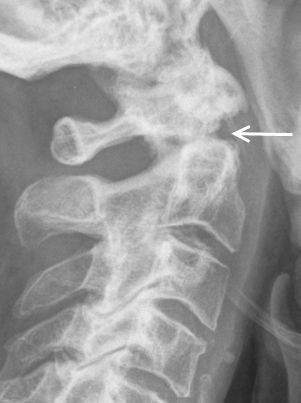
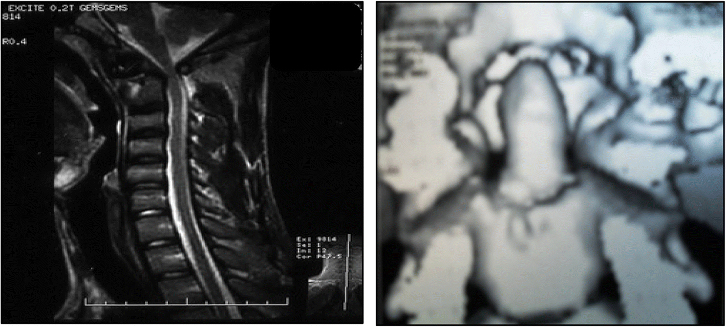
In such fractures, the lateral radiograph shows a break in the apparent ring that results from the superimposition of densities from the junction of pedicle and body, dens and body, and dorsal cortex of the C2 body. With a Type I fracture, the tip of the dens is broken. The type III fracture (or type 3 C2 body fracture) is a horizontally oriented, rostral fracture at the base of the dens. 2 and 3 associated with atlantoaxial instability were studied. doi:10.5435/00124635-201007000-00001 - Pubmed Figure 15 Type 3 Odontoid Fracture on CT coronal image. Physicians use a 3-type classification system to diagnose and treat odontoid fractures (Fig. Twelve patients with odontoid fracture type II, Figs.
Type 3 odontoid fracture update#
Odontoid Fractures: Update on Management. The Utility of MRI in the Evaluation of Odontoid Fractures. Span,applet,object,iframe,h1,h2,h3,h4,h5,h6,p,blockquote,pre,abbr,acronym,address,big,cite,code,del,dfn,em,ins,kbd,q,s,samp,small,strike,strong,sub,sup,tt,var,b,u,i,center,dl,dt,dd,ol,ul,li,fieldset,form,label,legend,table,caption,tbody,tfoot,thead,tr,th,td,article,aside,canvas,details,embed,figure,figcaption,header,hgroup,main,menu,nav,output,ruby,summary,time,mark,audio,video.


 0 kommentar(er)
